Projects on Fundamental Interactions on the Nanometer Scale
Our goal is to understand and predict TERS spectra by describing surface-immobilized molecules in the vicinity of plasmonic metal nanoparticles using computational quantum chemistry and classical electrodynamics. Combined, these theoretical methods shall deliver an explanation for specific Raman signatures of surface-immobilized molecules and a prediction for the limits of the lateral resolution under TERS conditions.
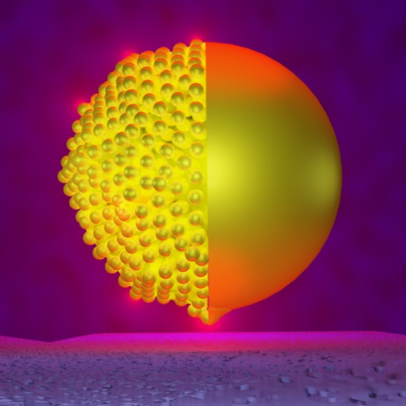 |
Computational ElectrodynamicsClassical electrodynamic methods, such as the Finite-Element-Method (FEM), are applied to different realistically shaped metal-nanoparticles (edges, vertices, defects etc.), in order to unravel their response to external laser light, i.e. the shape and confinement of the plasmonic field in the vicinity of the nanoparticle. These simulations produce field localizations that are able to explain observed lateral resolutions due to the locally confined electromagnetic response.
References
S. Trautmann, J. Aizpurua, I. Götz, A. Undisz, J. Dellith, H. Schneidewind, M. Rettenmayr, V. Deckert,
A classical description of subnanometer resolution by atomic features in metallic structures. Nanoscale, 2016, 8, 19, 10229. |
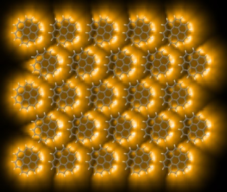 |
Quantum ChemistryHigh-level quantum chemical methods, such as DFT and TDDFT, are employed in order to study the local chemical effect of metal atoms and clusters on the polarizability of surface-immobilized molecules within a TERS setup. Current grid calculations already reveal possible sub-molecular resolution due to non-resonant chemical interactions alone.
References
F. Latorre, S. Kupfer, T. Bocklitz, D. Kinzel, S. Trautmann, S. Gräfe, V. Deckert,
Spatial resolution of tip-enhanced Raman spectroscopy - DFT assessment of the chemical effect. Nanoscale, 2017, 9, 1, 391. |
Experiments on High Lateral Resolution
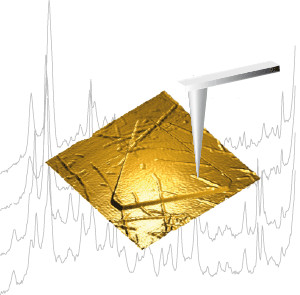 |
Direct experimental evidence of sub-nanometer resolutionSeveral TERS experiments on various bio-molecules reveal sub-nm resolution in accordance with the previous hypotheses. Systems investigated are protein fibrills and DNA strands. Interestingly such experiments can be done without any further “enhancing” techniques as gap-mode, resonance Raman etc., and even work under ambient conditions.
References
- Deckert-Gaudig, T.; Kurouski, D.; Hedegaard, M. A.; Singh, P.; Lednev, I. K.; Deckert, V.
Spatially resolved spectroscopic differentiation of hydrophilic and hydrophobic domains on individual insulin amyloid fibrils. Sci Rep., 2016, 6, 33575.
- Lin, X.-M.; Deckert-Gaudig, T.; Singh, P.; Siegmann, M.; Kupfer, S.; Zhang, Z.; et al.
Direct Base-to-Base Transitions in ssDNA Revealed by Tip-Enhanced Raman Scattering. arXiv.org, 2016, ,
- Deckert-Gaudig, T.; Kammer, E.; Deckert, V.
Tracking of nanoscale structural variations on a single amyloid fibril with tip-enhanced Raman scattering. J. Biophoton., 2012, 5, 3 , 215
|
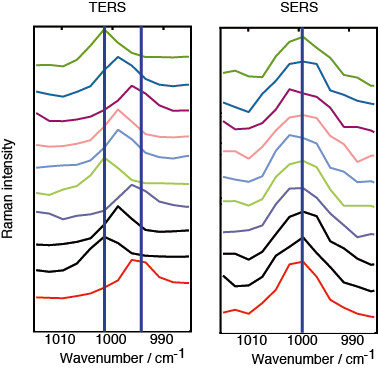
Indirect evidence …A different experimental approach evidencing high spatial resolution is based on the investigation of supposedly uniform monolayers. Here minute but statistically relevant spectral variations can be attributed to the presence of locally confined micro-ensembles, that do not yet show pronounced averaging effects. Hence, the detection volume must be confined to at most a few tens of molecules.
References
- Singh, P.; Deckert-Gaudig, T.; Schneidewind, H.; Kirsch, K.; van Schrojenstein Lantman, E.M.; Weckhuysen, B.M.; et al.
Differences in single and aggregated nanoparticle plasmon spectroscopy. Phys Chem Chem Phys., 2015, 17, 5, 2991.
|
 |